Please use this identifier to cite or link to this item:
http://hdl.handle.net/10267/33461| Title: | Network synchronization across the longitudinal axis of the developing rat hippocampus |
| Authors: | Siddiq, Bilal S. DaRosa, Andrew M. Evans, Madeline C. Gaudio, Elizabeth C. Mysiewicz, Steven C. Renna, Catelyn M. (Catie) Shamambo, Maleelo (Lelo) Velrajan, Srilakshmi (Sri) Van Vliet, Trevor Yu, Shuliang (Mogy) |
| Advisors: | Dougherty, Kelly A. |
| Keywords: | URCAS;Symposiums;Student research;2018 Spring;Class of 2020;Class of 2018;Class of 2019;Biology, Department of |
| Issue Date: | 27-Apr-2018 |
| Publisher: | Memphis, Tenn. : Rhodes College |
| Abstract: | Epilepsy is a neurological disorder characterized by recurrent seizures. Researching epilepsy requires animal models, as the brain manipulation required to understand epileptogenic activity is not possible in humans. Extracellular recordings measure network level activation in slice models of epilepsy. A magnesium-free solution is often used in slice physiology to increase excitation, through enhanced glutamate receptor activity. Magnesium blocks the activation of glutamate receptors via binding near the pore region. Magnesium also lowers calcium’s ability to initiate presynaptic neurotransmitter release. We aim to elucidate the mechanisms of a dorsal neuro-protective system, which reduces the excitability of dorsal hippocampal tissue with age. While the mechanisms underlying the magnesium-free model are well established, the effects of this model across the longitudinal axis of the hippocampus and throughout development have not been clearly characterized. We studied the effects of the magnesium-free model through extracellular field recordings of CA1 pyramidal neurons from dorsal and ventral hippocampal slices throughout development, and found young, dorsal hippocampal tissue to be the most excitable. Additionally, our data suggests the effect of no magnesium is minimal in ventral slices. These data support the theory of the dorsal protection system being critical in the reduction of excitability seen with age. |
| URI: | http://hdl.handle.net/10267/33461 |
| Appears in Collections: | Undergraduate Research and Creative Activity Symposium |
Files in This Item:
| File | Description | Size | Format | |
|---|---|---|---|---|
| 201804_Siddiq_Bilal_Poster_Slidedeck.pdf | 4.03 MB | Adobe PDF |  View/Open | |
| 201804_Siddiq_Bilal_SteadyState_Figure5.pdf | 193.5 kB | Adobe PDF | 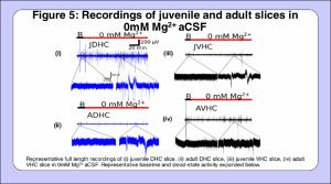 View/Open | |
| 201804_Siddiq_Bilal_Juvenile%3aAdultSlices_Figure8.pdf | 102.74 kB | Adobe PDF | 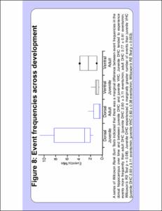 View/Open | |
| 201804_Siddiq_Bilal_IntericalEvents_Figure6.pdf | 107.94 kB | Adobe PDF | 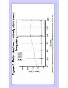 View/Open | |
| 201804_Siddiq_Bilal_Hippocampal Slice Physiology_Figure1.pdf | 300.61 kB | Adobe PDF | 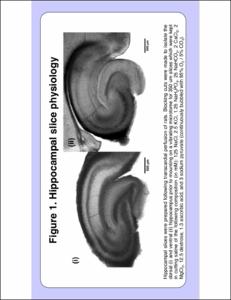 View/Open | |
| 201804_Siddiq_Bilal_FieldRecordingsinaCSF_Figure3.pdf | 183.98 kB | Adobe PDF | 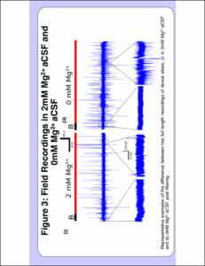 View/Open | |
| 201804_Siddiq_Bilal_Electrode placement and filtering_Figure2.pdf | 310.65 kB | Adobe PDF | 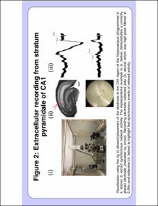 View/Open | |
| 201804_Siddiq_Bilal_EffectsofNoMag_Figure4.pdf | 218.38 kB | Adobe PDF | 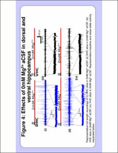 View/Open | |
| 201804_Siddiq_Bilal_EffectsinMag_Figure7.pdf | 96.06 kB | Adobe PDF | 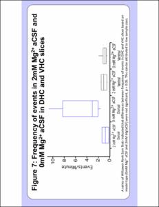 View/Open |
Items in DSpace are protected by copyright, with all rights reserved, unless otherwise indicated.